SUCLA2
Protein-coding gene in the species Homo sapiens
| SUCLA2 | |||||||||||||||||||||||||||||||||||||||||||||||||||
|---|---|---|---|---|---|---|---|---|---|---|---|---|---|---|---|---|---|---|---|---|---|---|---|---|---|---|---|---|---|---|---|---|---|---|---|---|---|---|---|---|---|---|---|---|---|---|---|---|---|---|---|
| Identifiers | |||||||||||||||||||||||||||||||||||||||||||||||||||
| Aliases | SUCLA2, A-BETA, MTDPS5, SCS-betaA, succinate-CoA ligase ADP-forming beta subunit, A-SCS, succinate-CoA ligase ADP-forming subunit beta, LINC00444 | ||||||||||||||||||||||||||||||||||||||||||||||||||
| External IDs | OMIM: 603921; MGI: 1306775; HomoloGene: 2856; GeneCards: SUCLA2; OMA:SUCLA2 - orthologs | ||||||||||||||||||||||||||||||||||||||||||||||||||
| |||||||||||||||||||||||||||||||||||||||||||||||||||
| |||||||||||||||||||||||||||||||||||||||||||||||||||
| |||||||||||||||||||||||||||||||||||||||||||||||||||
| |||||||||||||||||||||||||||||||||||||||||||||||||||
| |||||||||||||||||||||||||||||||||||||||||||||||||||
| Wikidata | |||||||||||||||||||||||||||||||||||||||||||||||||||
| |||||||||||||||||||||||||||||||||||||||||||||||||||


Succinyl-CoA ligase [ADP-forming] subunit beta, mitochondrial (SUCLA2), also known as ADP-forming succinyl-CoA synthetase (SCS-A), is an enzyme that in humans is encoded by the SUCLA2 gene on chromosome 13.[5][6][7]
Succinyl-CoA synthetase (SCS) is a mitochondrial matrix enzyme that acts as a heterodimer, composed of an invariant alpha subunit and a substrate-specific beta subunit. The protein encoded by this gene is an ATP-specific SCS beta subunit that dimerizes with the SCS alpha subunit to form SCS-A, an essential component of the tricarboxylic acid cycle. SCS-A hydrolyzes ATP to convert succinyl-CoA to succinate. Defects in this gene are a cause of myopathic mitochondrial DNA depletion syndrome. A pseudogene of this gene has been found on chromosome 6. [provided by RefSeq, Jul 2008][6]
Structure
SCS, also known as succinyl CoA ligase (SUCL), is a heterodimer composed of a catalytic α subunit encoded by the SUCLG1 gene and a β subunit encoded by either the SUCLA2 gene or the SUCLG2 gene, which determines the enzyme specificity for either ADP or GDP. SUCLA2 is the SCS variant containing the SUCLA2-encoded β subunit.[8][9][10] Amino acid sequence alignment of the two β subunit types reveals a homology of ~50% identity, with specific regions conserved throughout the sequences.[5]
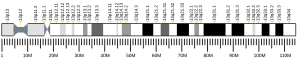
SUCLA2 is located on chromosome 13 and contains 13 exons.[6]
Function
As a subunit of SCS, SUCLA2 is a mitochondrial matrix enzyme that catalyzes the reversible conversion of succinyl-CoA to succinate and Acetoacetyl CoA, accompanied by the substrate-level phosphorylation of ADP to ATP, as a step in the tricarboxylic acid (TCA) cycle.[8][9][10] The ATP generated is then consumed in catabolic pathways.[9] Since substrate-level phosphorylation does not require oxygen for ATP production, this reaction can rescue cells from cytosolic ATP depletion during ischemia.[10] The reverse reaction generates succinyl-CoA from succinate to fuel ketone body and heme synthesis.[8][10]
While SCS is ubiquitously expressed, SUCLA2 is predominantly expressed in catabolic tissues reliant on ATP as their main energy source, including the heart, brain, and skeletal muscle.[5][7][10] Within the brain, SUCLA2 is found exclusively in neurons; meanwhile, both SUCLA2 and SUCLG2 are absent in astrocytes, microglia, and oligodendrocytes. In order to acquire succinate to continue the TCA cycle, these cells may instead synthesize succinate through GABAmetabolism of α-ketoglutarate or ketone body metabolism of succinyl-CoA.[9][10]
Clinical significance
Mutations in the SUCLA2 gene are associated with mitochondrial DNA (mtDNA) depletion syndrome.[11][12] Symptoms include early-onset low muscle tone, severe muscular atrophy, scoliosis, movement disorders such as dystonia and hyperkinesia, epilepsy, and growth retardation. Because succinic acid cannot be made from succinyl coa, treatment is with oral succinic acid, which allows the Krebs cycle and electron transport chain to function correctly. Other treatments for managing symptoms include exercises to promote mobility and respiratory assistance, baclofen to treat dystonia and hyperkinesia, and antiepileptic drugs for seizures.[11][13]
There is a relatively high incidence of a specific SUCLA2 mutation in the Faroe Islands due to a founder effect. This particular mutation is often associated with early lethality.[14] Two additional founder mutations have been discovered in the Scandinavian population, in addition to the known SUCLA2 founder mutation in the Faroe Islands.[15] These patients show a higher variability in outcomes with several patients with SUCLA2 missense mutation surviving into adulthood. This variability suggests that SUCLA2 missense mutations may be associated with residual enzyme activity.[15]
Coenzyme Q10 and antioxidants have been used to treat mitochondrial DNA depletion syndrome, but there is currently no evidence that these treatments result in clinical benefit.[13][16]
Mutations in the SUCLA2 gene leading to SUCLA2 deficiency result in Leigh's or a Leigh-like syndrome with the onset of severe hypotonia, muscular atrophy, sensorineural hearing impairment, and often death in early childhood.[8][10]
See also
References
- ^ a b c GRCh38: Ensembl release 89: ENSG00000136143 – Ensembl, May 2017
- ^ a b c GRCm38: Ensembl release 89: ENSMUSG00000022110 – Ensembl, May 2017
- ^ "Human PubMed Reference:". National Center for Biotechnology Information, U.S. National Library of Medicine.
- ^ "Mouse PubMed Reference:". National Center for Biotechnology Information, U.S. National Library of Medicine.
- ^ a b c Johnson JD, Mehus JG, Tews K, Milavetz BI, Lambeth DO (October 1998). "Genetic evidence for the expression of ATP- and GTP-specific succinyl-CoA synthetases in multicellular eucaryotes". The Journal of Biological Chemistry. 273 (42): 27580–6. doi:10.1074/jbc.273.42.27580. PMID 9765291.
- ^ a b c "Entrez Gene: SUCLA2 succinate-CoA ligase, ADP-forming, beta subunit".
- ^ a b Matilainen S, Isohanni P, Euro L, Lönnqvist T, Pihko H, Kivelä T, Knuutila S, Suomalainen A (March 2015). "Mitochondrial encephalomyopathy and retinoblastoma explained by compound heterozygosity of SUCLA2 point mutation and 13q14 deletion". European Journal of Human Genetics. 23 (3): 325–30. doi:10.1038/ejhg.2014.128. PMC 4326715. PMID 24986829.
- ^ a b c d Miller C, Wang L, Ostergaard E, Dan P, Saada A (May 2011). "The interplay between SUCLA2, SUCLG2, and mitochondrial DNA depletion" (PDF). Biochimica et Biophysica Acta (BBA) - Molecular Basis of Disease. 1812 (5): 625–9. doi:10.1016/j.bbadis.2011.01.013. PMID 21295139.
- ^ a b c d Dobolyi A, Bagó AG, Gál A, Molnár MJ, Palkovits M, Adam-Vizi V, Chinopoulos C (April 2015). "Localization of SUCLA2 and SUCLG2 subunits of succinyl CoA ligase within the cerebral cortex suggests the absence of matrix substrate-level phosphorylation in glial cells of the human brain" (PDF). Journal of Bioenergetics and Biomembranes. 47 (1–2): 33–41. doi:10.1007/s10863-014-9586-4. PMID 25370487. S2CID 41101828.
- ^ a b c d e f g Dobolyi A, Ostergaard E, Bagó AG, Dóczi T, Palkovits M, Gál A, Molnár MJ, Adam-Vizi V, Chinopoulos C (January 2015). "Exclusive neuronal expression of SUCLA2 in the human brain" (PDF). Brain Structure & Function. 220 (1): 135–51. doi:10.1007/s00429-013-0643-2. PMID 24085565. S2CID 105582.
- ^ a b Ostergaard E (May 2009). "SUCLA2-Related Mitochondrial DNA Depletion Syndrome, Encephalomyopathic Form, with Mild Methylmalonic Acuduria". In Pagon RA, Adam MP, Ardinger HH, Wallace SE, Amemiya A, Bean LJ, Bird TD, Fong CT, Mefford HC, Smith RJ, Stephens K (eds.). GeneReviews [Internet]. Seattle: University of Washington, Seattle. PMID 20301762.
- ^ El-Hattab AW, Scaglia F (April 2013). "Mitochondrial DNA depletion syndromes: review and updates of genetic basis, manifestations, and therapeutic options". review. Neurotherapeutics. 10 (2): 186–98. doi:10.1007/s13311-013-0177-6. PMC 3625391. PMID 23385875.
- ^ a b Parikh S, Saneto R, Falk MJ, Anselm I, Cohen BH, Haas R, Medicine Society TM (November 2009). "A modern approach to the treatment of mitochondrial disease". primary source. Current Treatment Options in Neurology. 11 (6): 414–30. doi:10.1007/s11940-009-0046-0. PMC 3561461. PMID 19891905.
- ^ Ostergaard E, Hansen FJ, Sorensen N, Duno M, Vissing J, Larsen PL, Faeroe O, Thorgrimsson S, Wibrand F, Christensen E, Schwartz M (March 2007). "Mitochondrial encephalomyopathy with elevated methylmalonic acid is caused by SUCLA2 mutations". primary source. Brain. 130 (Pt 3): 853–61. CiteSeerX 10.1.1.321.3705. doi:10.1093/brain/awl383. PMID 17287286.
- ^ a b Carrozzo R, Verrigni D, Rasmussen M, de Coo R, Amartino H, Bianchi M, Buhas D, Mesli S, Naess K, Born AP, Woldseth B, Prontera P, Batbayli M, Ravn K, Joensen F, Cordelli DM, Santorelli FM, Tulinius M, Darin N, Duno M, Jouvencel P, Burlina A, Stangoni G, Bertini E, Redonnet-Vernhet I, Wibrand F, Dionisi-Vici C, Uusimaa J, Vieira P, Osorio AN, McFarland R, Taylor RW, Holme E, Ostergaard E (March 2016). "Succinate-CoA ligase deficiency due to mutations in SUCLA2 and SUCLG1: phenotype and genotype correlations in 71 patients". primary source. Journal of Inherited Metabolic Disease. 39 (2): 243–52. doi:10.1007/s10545-015-9894-9. PMID 26475597. S2CID 7881205.
- ^ Pfeffer G, Majamaa K, Turnbull DM, Thorburn D, Chinnery PF (April 2012). "Treatment for mitochondrial disorders". review. The Cochrane Database of Systematic Reviews. 4 (4): CD004426. doi:10.1002/14651858.CD004426.pub3. PMC 7201312. PMID 22513923.
Further reading
- Maruyama K, Sugano S (January 1994). "Oligo-capping: a simple method to replace the cap structure of eukaryotic mRNAs with oligoribonucleotides". Gene. 138 (1–2): 171–4. doi:10.1016/0378-1119(94)90802-8. PMID 8125298.
- Suzuki Y, Yoshitomo-Nakagawa K, Maruyama K, Suyama A, Sugano S (October 1997). "Construction and characterization of a full length-enriched and a 5'-end-enriched cDNA library". Gene. 200 (1–2): 149–56. doi:10.1016/S0378-1119(97)00411-3. PMID 9373149.
- Scanlan MJ, Gordan JD, Williamson B, Stockert E, Bander NH, Jongeneel V, Gure AO, Jäger D, Jäger E, Knuth A, Chen YT, Old LJ (November 1999). "Antigens recognized by autologous antibody in patients with renal-cell carcinoma". International Journal of Cancer. 83 (4): 456–64. doi:10.1002/(SICI)1097-0215(19991112)83:4<456::AID-IJC4>3.0.CO;2-5. PMID 10508479. S2CID 21839750.
- Furuyama K, Sassa S (March 2000). "Interaction between succinyl CoA synthetase and the heme-biosynthetic enzyme ALAS-E is disrupted in sideroblastic anemia". The Journal of Clinical Investigation. 105 (6): 757–64. doi:10.1172/JCI6816. PMC 377455. PMID 10727444.
- Cox TC, Sadlon TJ, Schwarz QP, Matthews CS, Wise PD, Cox LL, Bottomley SS, May BK (February 2004). "The major splice variant of human 5-aminolevulinate synthase-2 contributes significantly to erythroid heme biosynthesis". The International Journal of Biochemistry & Cell Biology. 36 (2): 281–95. doi:10.1016/S1357-2725(03)00246-2. PMID 14643893.
- Rush J, Moritz A, Lee KA, Guo A, Goss VL, Spek EJ, Zhang H, Zha XM, Polakiewicz RD, Comb MJ (January 2005). "Immunoaffinity profiling of tyrosine phosphorylation in cancer cells". Nature Biotechnology. 23 (1): 94–101. doi:10.1038/nbt1046. PMID 15592455. S2CID 7200157.
- Elpeleg O, Miller C, Hershkovitz E, Bitner-Glindzicz M, Bondi-Rubinstein G, Rahman S, Pagnamenta A, Eshhar S, Saada A (June 2005). "Deficiency of the ADP-forming succinyl-CoA synthase activity is associated with encephalomyopathy and mitochondrial DNA depletion". American Journal of Human Genetics. 76 (6): 1081–6. doi:10.1086/430843. PMC 1196446. PMID 15877282.
- Rual JF, Venkatesan K, Hao T, Hirozane-Kishikawa T, Dricot A, Li N, Berriz GF, Gibbons FD, Dreze M, Ayivi-Guedehoussou N, Klitgord N, Simon C, Boxem M, Milstein S, Rosenberg J, Goldberg DS, Zhang LV, Wong SL, Franklin G, Li S, Albala JS, Lim J, Fraughton C, Llamosas E, Cevik S, Bex C, Lamesch P, Sikorski RS, Vandenhaute J, Zoghbi HY, Smolyar A, Bosak S, Sequerra R, Doucette-Stamm L, Cusick ME, Hill DE, Roth FP, Vidal M (October 2005). "Towards a proteome-scale map of the human protein-protein interaction network". Nature. 437 (7062): 1173–8. Bibcode:2005Natur.437.1173R. doi:10.1038/nature04209. PMID 16189514. S2CID 4427026.
External links
- GeneReview/NCBI/NIH/UW entry on SUCLA2-Related Mitochondrial DNA Depletion Syndrome, Encephalomyopathic Form, with Mild Methylmalonic Aciduria


