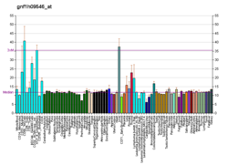
| CDK3 |
|---|
|
| PDBに登録されている構造 |
|---|
| PDB | Human UniProt検索: RCSB PDBe PDBj |
|---|
|
|
| 識別子 |
|---|
| 記号 | CDK3, cyclin dependent kinase 3 |
|---|
| 外部ID | OMIM: 123828 HomoloGene: 74387 GeneCards: CDK3 |
|---|
| 遺伝子の位置 (ヒト) |
|---|
 | | 染色体 | 17番染色体 (ヒト)[1] |
|---|
| | バンド | データ無し | 開始点 | 76,000,855 bp[1] |
|---|
| 終点 | 76,005,999 bp[1] |
|---|
|
|
| 遺伝子オントロジー |
|---|
| 分子機能 | • protein serine/threonine kinase activity
• ATP binding
• キナーゼ活性
• ヌクレオチド結合
• protein kinase activity
• トランスフェラーゼ活性
• cyclin-dependent protein serine/threonine kinase activity
• 血漿タンパク結合
• cyclin binding
|
|---|
| 細胞の構成要素 | • cyclin-dependent protein kinase holoenzyme complex
• 細胞核
• 細胞質
|
|---|
| 生物学的プロセス | • 細胞分裂
• 細胞増殖
• 細胞周期
• G0 to G1 transition
• cellular response to DNA damage stimulus
• リン酸化
• タンパク質リン酸化
• シグナル伝達
• 多細胞個体の発生
• positive regulation of cell population proliferation
• 有機物への反応
• regulation of G2/M transition of mitotic cell cycle
• 遺伝子発現調節
• G1/S transition of mitotic cell cycle
• regulation of cell cycle
|
|---|
| 出典:Amigo / QuickGO |
|
| オルソログ |
|---|
| 種 | ヒト | マウス |
|---|
| Entrez | | |
|---|
| Ensembl | | |
|---|
| UniProt | | |
|---|
RefSeq
(mRNA) | | |
|---|
RefSeq
(タンパク質) | | |
|---|
場所
(UCSC) | Chr 17: 76 – 76.01 Mb | n/a |
|---|
| PubMed検索 | [2] | n/a |
|---|
| ウィキデータ |
|
サイクリン依存性キナーゼ3(サイクリンいぞんせいキナーゼ3、英: cyclin-dependent kinase 3、略称: CDK3)は、ヒトではCDK3遺伝子によってコードされる酵素である[3][4]。
機能
CDK3遺伝子はサイクリン依存性キナーゼ(CDK)ファミリーのメンバーであるCDK3タンパク質をコードする。CDK3タンパク質はS期への移行を促進するが、その一部はE2Fファミリーの転写因子の活性化によるものである。また、サイクリンCと結合してRbタンパク質をリン酸化し、G0期からの脱出を促進する[4]。
出典
- ^ a b c GRCh38: Ensembl release 89: ENSG00000250506 - Ensembl, May 2017
- ^ Human PubMed Reference:
- ^ “A family of human cdc2-related protein kinases”. EMBO J 11 (8): 2909–17. (Aug 1992). doi:10.1002/j.1460-2075.1992.tb05360.x. PMC 556772. PMID 1639063. https://www.ncbi.nlm.nih.gov/pmc/articles/PMC556772/.
- ^ a b “Entrez Gene: CDK3 cyclin-dependent kinase 3”. 2022年3月5日閲覧。
関連文献
- “Chromosomal mapping of members of the cdc2 family of protein kinases, cdk3, cdk6, PISSLRE, and PITALRE, and a cdk inhibitor, p27Kip1, to regions involved in human cancer.”. Cancer Res. 55 (6): 1199–205. (1995). PMID 7882308.
- “Cdi1, a human G1 and S phase protein phosphatase that associates with Cdk2.”. Cell 75 (4): 791–803. (1993). doi:10.1016/0092-8674(93)90498-F. PMID 8242750.
- “Phorbol ester inhibits the phosphorylation of the retinoblastoma protein without suppressing cyclin D-associated kinase in vascular smooth muscle cells.”. J. Biol. Chem. 271 (14): 8345–51. (1996). doi:10.1074/jbc.271.14.8345. PMID 8626531.
- “Suppression of apoptosis by dominant negative mutants of cyclin-dependent protein kinases.”. J. Biol. Chem. 271 (17): 10205–9. (1996). doi:10.1074/jbc.271.17.10205. PMID 8626584.
- “Differential effects of cdk2 and cdk3 on the control of pRb and E2F function during G1 exit.”. Genes Dev. 10 (7): 851–61. (1996). doi:10.1101/gad.10.7.851. PMID 8846921.
- “Interaction between Cdc37 and Cdk4 in human cells.”. Oncogene 14 (16): 1999–2004. (1997). doi:10.1038/sj.onc.1201036. PMID 9150368.
- “Investigation of the cell cycle regulation of cdk3-associated kinase activity and the role of cdk3 in proliferation and transformation.”. Oncogene 17 (17): 2259–69. (1998). doi:10.1038/sj.onc.1202145. PMID 9811456.
- “ik3-1/Cables is a substrate for cyclin-dependent kinase 3 (cdk 3).”. Eur. J. Biochem. 268 (23): 6076–82. (2002). doi:10.1046/j.0014-2956.2001.02555.x. PMID 11733001.
- “ik3-2, a relative to ik3-1/cables, is associated with cdk3, cdk5, and c-abl.”. Biochim. Biophys. Acta 1574 (2): 157–63. (2002). doi:10.1016/S0167-4781(01)00367-0. PMID 11955625.
- “Explant-induced reactivation of herpes simplex virus occurs in neurons expressing nuclear cdk2 and cdk4”. J. Virol. 76 (15): 7724–35. (2002). doi:10.1128/JVI.76.15.7724-7735.2002. PMC 136347. PMID 12097586. https://www.ncbi.nlm.nih.gov/pmc/articles/PMC136347/.
- “Complete sequencing and characterization of 21,243 full-length human cDNAs”. Nat. Genet. 36 (1): 40–5. (2004). doi:10.1038/ng1285. PMID 14702039.
- “Cyclin C/cdk3 promotes Rb-dependent G0 exit”. Cell 117 (2): 239–51. (2004). doi:10.1016/S0092-8674(04)00300-9. PMID 15084261.
- “Time-resolved mass spectrometry of tyrosine phosphorylation sites in the epidermal growth factor receptor signaling network reveals dynamic modules”. Mol. Cell. Proteomics 4 (9): 1240–50. (2005). doi:10.1074/mcp.M500089-MCP200. PMID 15951569.
- “A probability-based approach for high-throughput protein phosphorylation analysis and site localization”. Nat. Biotechnol. 24 (10): 1285–92. (2006). doi:10.1038/nbt1240. PMID 16964243.
- “Global, in vivo, and site-specific phosphorylation dynamics in signaling networks”. Cell 127 (3): 635–48. (2006). doi:10.1016/j.cell.2006.09.026. PMID 17081983.
- “Proteomics analysis of protein kinases by target class-selective prefractionation and tandem mass spectrometry”. Mol. Cell. Proteomics 6 (3): 537–47. (2007). doi:10.1074/mcp.T600062-MCP200. PMID 17192257. https://hzi.openrepository.com/bitstream/10033/19756/1/Wissing%20et%20al_final.pdf.
外部リンク


