S100A6
Human protein and coding gene
| S100A6 | |||||||||||||||||||||||||||||||||||||||||||||||||||
|---|---|---|---|---|---|---|---|---|---|---|---|---|---|---|---|---|---|---|---|---|---|---|---|---|---|---|---|---|---|---|---|---|---|---|---|---|---|---|---|---|---|---|---|---|---|---|---|---|---|---|---|
 | |||||||||||||||||||||||||||||||||||||||||||||||||||
| Identifiers | |||||||||||||||||||||||||||||||||||||||||||||||||||
| Aliases | S100A6, 2A9, 5B10, CABP, CACY, PRA, S100 calcium binding protein A6, S10A6 | ||||||||||||||||||||||||||||||||||||||||||||||||||
| External IDs | OMIM: 114110; MGI: 1339467; HomoloGene: 7925; GeneCards: S100A6; OMA:S100A6 - orthologs | ||||||||||||||||||||||||||||||||||||||||||||||||||
| |||||||||||||||||||||||||||||||||||||||||||||||||||
| |||||||||||||||||||||||||||||||||||||||||||||||||||
| |||||||||||||||||||||||||||||||||||||||||||||||||||
| |||||||||||||||||||||||||||||||||||||||||||||||||||
| |||||||||||||||||||||||||||||||||||||||||||||||||||
| Wikidata | |||||||||||||||||||||||||||||||||||||||||||||||||||
| |||||||||||||||||||||||||||||||||||||||||||||||||||


S100 calcium-binding protein A6 (S100A6) is a protein that in humans is encoded by the S100A6 gene.[5]
Function
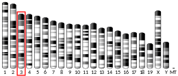
The protein encoded by this gene is a member of the S100 family of proteins containing 2 EF-hand calcium-binding motifs. S100 proteins are localized in the cytoplasm and/or nucleus of a wide range of cells, and involved in the regulation of a number of cellular processes such as cell cycle progression and differentiation. S100 genes include at least 13 members which are located as a cluster on chromosome 1q21. This protein may function in stimulation of Ca2+-dependent insulin release, stimulation of prolactin secretion, and exocytosis. Chromosomal rearrangements and altered expression of this gene have been implicated in melanoma.[5]
Interactions
S100A6 has been shown to interact with S100B[6][7] and SUGT1.[8]
Pathology
S100A6 to be reported as possible diagnostic marker of papillary thyroid carcinoma.[9]
References
- ^ a b c GRCh38: Ensembl release 89: ENSG00000197956 – Ensembl, May 2017
- ^ a b c GRCm38: Ensembl release 89: ENSMUSG00000001025 – Ensembl, May 2017
- ^ "Human PubMed Reference:". National Center for Biotechnology Information, U.S. National Library of Medicine.
- ^ "Mouse PubMed Reference:". National Center for Biotechnology Information, U.S. National Library of Medicine.
- ^ a b "Entrez Gene: S100A6 S100 calcium binding protein A6".
- ^ Deloulme JC, Assard N, Mbele GO, Mangin C, Kuwano R, Baudier J (November 2000). "S100A6 and S100A11 are specific targets of the calcium- and zinc-binding S100B protein in vivo". J. Biol. Chem. 275 (45): 35302–10. doi:10.1074/jbc.M003943200. PMID 10913138.
- ^ Yang Q, O'Hanlon D, Heizmann CW, Marks A (February 1999). "Demonstration of heterodimer formation between S100B and S100A6 in the yeast two-hybrid system and human melanoma". Exp. Cell Res. 246 (2): 501–9. doi:10.1006/excr.1998.4314. PMID 9925766.
- ^ Nowotny M, Spiechowicz M, Jastrzebska B, Filipek A, Kitagawa K, Kuznicki J (July 2003). "Calcium-regulated interaction of Sgt1 with S100A6 (calcyclin) and other S100 proteins". J. Biol. Chem. 278 (29): 26923–8. doi:10.1074/jbc.M211518200. PMID 12746458.
- ^ Sofiadis A, Dinets A, Orre LM, Branca RM, Juhlin CC, Foukakis T, Wallin G, Höög A, Hulchiy M, Zedenius J, Larsson C, Lehtiö J (October 2010). "Proteomic study of thyroid tumors reveals frequent up-regulation of the Ca2+ -binding protein S100A6 in papillary thyroid carcinoma". Thyroid. 20 (10): 1067–76. doi:10.1089/thy.2009.0400. PMID 20629554.
Further reading
- Schäfer BW, Heizmann CW (1996). "The S100 family of EF-hand calcium-binding proteins: functions and pathology". Trends Biochem. Sci. 21 (4): 134–40. doi:10.1016/S0968-0004(96)80167-8. PMID 8701470.
- Minami H, Tokumitsu H, Mizutani A, Watanabe Y, Watanabe M, Hidaka H (1992). "Specific binding of CAP-50 to calcyclin". FEBS Lett. 305 (3): 217–9. doi:10.1016/0014-5793(92)80671-3. PMID 1299619.
- Engelkamp D, Schäfer BW, Erne P, Heizmann CW (1992). "S100 alpha, CAPL, and CACY: molecular cloning and expression analysis of three calcium-binding proteins from human heart". Biochemistry. 31 (42): 10258–64. doi:10.1021/bi00157a012. PMID 1384693.
- Tomida Y, Terasawa M, Kobayashi R, Hidaka H (1992). "Calcyclin and calvasculin exist in human platelets". Biochem. Biophys. Res. Commun. 189 (3): 1310–6. doi:10.1016/0006-291X(92)90216-8. PMID 1482346.
- Filipek A, Gerke V, Weber K, Kuźnicki J (1991). "Characterization of the cell-cycle-regulated protein calcyclin from Ehrlich ascites tumor cells. Identification of two binding proteins obtained by Ca2(+)-dependent affinity chromatography". Eur. J. Biochem. 195 (3): 795–800. doi:10.1111/j.1432-1033.1991.tb15768.x. PMID 1999197.
- Murphy LC, Murphy LJ, Tsuyuki D, Duckworth ML, Shiu RP (1988). "Cloning and characterization of a cDNA encoding a highly conserved, putative calcium binding protein, identified by an anti-prolactin receptor antiserum". J. Biol. Chem. 263 (5): 2397–401. doi:10.1016/S0021-9258(18)69220-8. PMID 2448309.
- Gabius HJ, Bardosi A, Gabius S, Hellmann KP, Karas M, Kratzin H (1989). "Identification of a cell cycle-dependent gene product as a sialic acid-binding protein". Biochem. Biophys. Res. Commun. 163 (1): 506–12. doi:10.1016/0006-291X(89)92166-9. PMID 2775283.
- Ferrari S, Calabretta B, deRiel JK, Battini R, Ghezzo F, Lauret E, Griffin C, Emanuel BS, Gurrieri F, Baserga R (1987). "Structural and functional analysis of a growth-regulated gene, the human calcyclin". J. Biol. Chem. 262 (17): 8325–32. doi:10.1016/S0021-9258(18)47567-9. hdl:11380/811306. PMID 3036810.
- Calabretta B, Battini R, Kaczmarek L, de Riel JK, Baserga R (1986). "Molecular cloning of the cDNA for a growth factor-inducible gene with strong homology to S-100, a calcium-binding protein". J. Biol. Chem. 261 (27): 12628–32. doi:10.1016/S0021-9258(18)67137-6. hdl:11380/811299. PMID 3755724.
- Schäfer BW, Wicki R, Engelkamp D, Mattei MG, Heizmann CW (1995). "Isolation of a YAC clone covering a cluster of nine S100 genes on human chromosome 1q21: rationale for a new nomenclature of the S100 calcium-binding protein family". Genomics. 25 (3): 638–43. doi:10.1016/0888-7543(95)80005-7. PMID 7759097.
- Engelkamp D, Schäfer BW, Mattei MG, Erne P, Heizmann CW (1993). "Six S100 genes are clustered on human chromosome 1q21: identification of two genes coding for the two previously unreported calcium-binding proteins S100D and S100E". Proc. Natl. Acad. Sci. U.S.A. 90 (14): 6547–51. Bibcode:1993PNAS...90.6547E. doi:10.1073/pnas.90.14.6547. PMC 46969. PMID 8341667.
- Filipek A, Wojda U (1996). "p30, a novel protein target of mouse calcyclin (S100A6)". Biochem. J. 320 ( Pt 2) (Pt 2): 585–7. doi:10.1042/bj3200585. PMC 1217969. PMID 8973570.
- Kordowska J, Stafford WF, Wang CL (1998). "Ca2+ and Zn2+ bind to different sites and induce different conformational changes in human calcyclin". Eur. J. Biochem. 253 (1): 57–66. doi:10.1046/j.1432-1327.1998.2530057.x. PMID 9578461.
- Yang Q, O'Hanlon D, Heizmann CW, Marks A (1999). "Demonstration of heterodimer formation between S100B and S100A6 in the yeast two-hybrid system and human melanoma". Exp. Cell Res. 246 (2): 501–9. doi:10.1006/excr.1998.4314. PMID 9925766.
- Sudo T, Hidaka H (1999). "Characterization of the calcyclin (S100A6) binding site of annexin XI-A by site-directed mutagenesis". FEBS Lett. 444 (1): 11–4. doi:10.1016/S0014-5793(99)00014-9. PMID 10037139.
- Deloulme JC, Assard N, Mbele GO, Mangin C, Kuwano R, Baudier J (2000). "S100A6 and S100A11 are specific targets of the calcium- and zinc-binding S100B protein in vivo". J. Biol. Chem. 275 (45): 35302–10. doi:10.1074/jbc.M003943200. PMID 10913138.
- Li Y, Yang L, Cui JT, Li WM, Guo RF, Lu YY (2002). "Construction of cDNA representational difference analysis based on two cDNA libraries and identification of garlic inducible expression genes in human gastric cancer cells". World J. Gastroenterol. 8 (2): 208–12. doi:10.3748/wjg.v8.i2.208. PMC 4658352. PMID 11925593.
- Otterbein LR, Kordowska J, Witte-Hoffmann C, Wang CL, Dominguez R (2002). "Crystal structures of S100A6 in the Ca(2+)-free and Ca(2+)-bound states: the calcium sensor mechanism of S100 proteins revealed at atomic resolution". Structure. 10 (4): 557–67. doi:10.1016/S0969-2126(02)00740-2. PMID 11937060.
- Filipek A, Jastrzebska B, Nowotny M, Kuznicki J (2002). "CacyBP/SIP, a calcyclin and Siah-1-interacting protein, binds EF-hand proteins of the S100 family". J. Biol. Chem. 277 (32): 28848–52. doi:10.1074/jbc.M203602200. PMID 12042313.
- v
- t
- e
PDB gallery
-
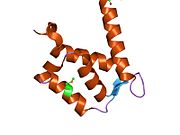
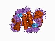 1a03: THE THREE-DIMENSIONAL STRUCTURE OF CA2+-BOUND CALCYCLIN: IMPLICATIONS FOR CA2+-SIGNAL TRANSDUCTION BY S100 PROTEINS, NMR, 20 STRUCTURES
 -
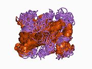 1cnp: THE STRUCTURE OF CALCYCLIN REVEALS A NOVEL HOMODIMERIC FOLD FOR S100 CA2+-BINDING PROTEINS, NMR, 22 STRUCTURES
 -
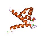
 1jwd: Ca2+-induced Structural Changes in Calcyclin: High-resolution Solution Structure of Ca2+-bound Calcyclin.
 -
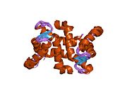
 1k8u: CRYSTAL STRUCTURE OF CALCIUM-FREE (OR APO) HUMAN S100A6; CYS3MET MUTANT (SELENOMETHIONINE DERIVATIVE)
 -
 1k96: CRYSTAL STRUCTURE OF CALCIUM BOUND HUMAN S100A6
 -
 1k9k: CRYSTAL STRUCTURE OF CALCIUM BOUND HUMAN S100A6
 -
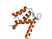 1k9p: CRYSTAL STRUCTURE OF CALCIUM FREE (OR APO) HUMAN S100A6
 -
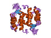 2cnp: HIGH RESOLUTION SOLUTION STRUCTURE OF APO RABBIT CALCYCLIN, NMR, 22 STRUCTURES


 | This article on a gene on human chromosome 1 is a stub. You can help Wikipedia by expanding it. |
- v
- t
- e


